Engineers are available to assist.


Flow Cytometry is an analytical technique used in a variety of life science applications for counting, inspecting or sorting particles in solution such as single cells. This technique enables the analysis of mixed cell populations - for example from blood, bone marrow, or even from solid tissues such as tumours when these are dissociated into single cells. This allows for the quick and accurate diagnosis of a variety of diseases or sorting of cells for further analysis. Flow cytometry is used in a variety of disciplines such as immunology, cancer, virology and molecular biology, as well as infectious disease monitoring.
Flow cytometers are diagnostic devices that use optofluidic systems in which one or multiple lasers are focused onto the sample being analysed as particles flow past. This produces either scattered or fluorescent light signals depending on the particle characteristics: particle shape, size or dye used for staining. These signals then pass through filters to the detector(s) - typically photodiodes or photomultiplier tubes. The beam path of these systems usually consists of lenses, optical filters, mirrors, prisms and other optical components to direct the light. The optical components involved are crucial in improving the accuracy of these systems and successfully delivering the signal from the flow cell to the detector(s).

Flow cytometry is the primary technology for inspection and detection of blood and other bodily fluid ailments. A flow cytometer is made up of three critical sub systems – a fluidics system, an electronic detection system, and an optical system.
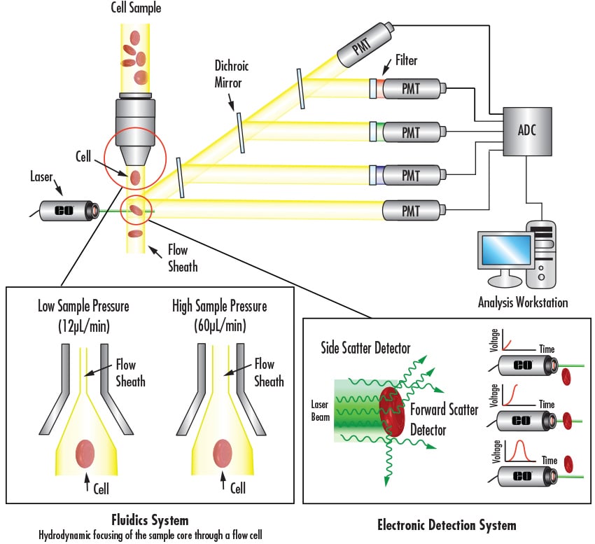
High flow rates used for qualitative measurements:
Lower flow rates used for higher resolution:
Forward-scattered light: measurement of diffracted light slightly off-axis of the laser beam, which detects particles within a given size range
Side-scattered light: measurement of mostly refracted and reflected light at any interface in cell where refractive index changes that is proportional to cell complexity and granularity
Equipment
Excitation Optics: laser and lenses for shaping and focusing of laser beam
Emission Optics: various lenses to collect scatter and mirrors, filters, and beamsplitters for proper routing
Learn more about the how fluorophores and optical filters work for flourecsence microsocpy.
Many techniques and methods are used in order to view, diagnose, and treat blood and other bodily fluids. The most common techniques include Flow Cytometry, Cell Sorting, Optofluidics, and Microscopy.
Fluorescence activated cell sorting (FACS) is a specific branch of flow cytometry that actively sorts a heterogeneous collection of cells into various containers a single cell at a time. This is done using general light scattering and fluorescence principles based off of each cell’s characteristics.
Technology that combines the field of microfluidics with optics. The primary applications include broad covering liquid displays, energy, and optical lenses, but the primary startup company drive is focusing on lab-on-chip devices, biosensors, and molecular imaging systems.
A powerful drug discovery process used heavily in pharmaceuticals. Typically an automated procedure that allows for quicker deployment of novel drugs with less risk for human error.
Traditional light microscopes are used to view histology slides or prepared cells and samples. Higher end microscopes known as confocal or multiphoton microscopes utilize multiple lasers, scanning mirrors, motorized actuators, and an array of high end detectors to better understand intracellular activity or protein-protein interactions.
A specialized type of flow cytometry that uses the fluorescent and scattering characteristics of biological cells to sort them into separate containers. It is used for separating in a heterogeneous mixture one at a time.

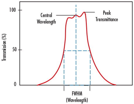
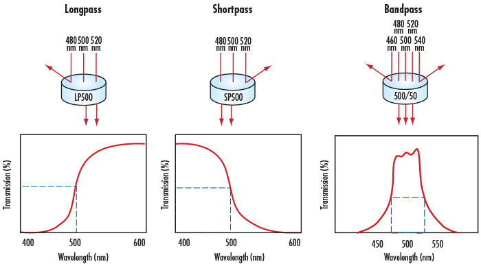
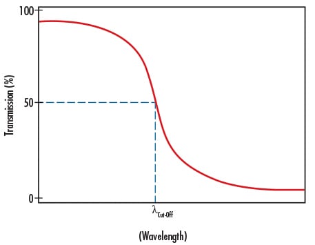
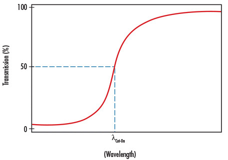
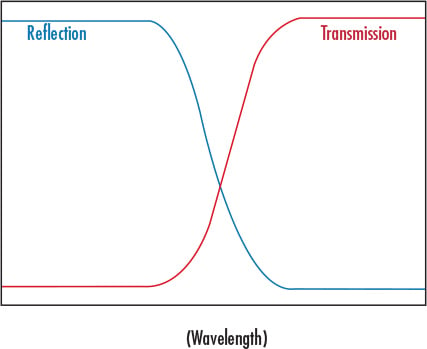
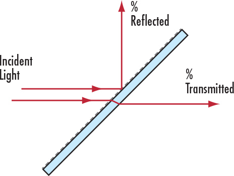
Optics are crucial to many life sciences applications and medical devices including flow cytometers. Beamsplitters and various types of filters, such as bandpass, dichroic, longpass, and shortpass filters, are only a few of the most prominently utilized.
Below are common ailments of the blood that are detected by advanced diagnostic techniques such as flow cytometry. Optical advancements allow these ailments to be more easily detected and treated, providing a means for making medical technology and equipment more quick, portable, and simple to use.
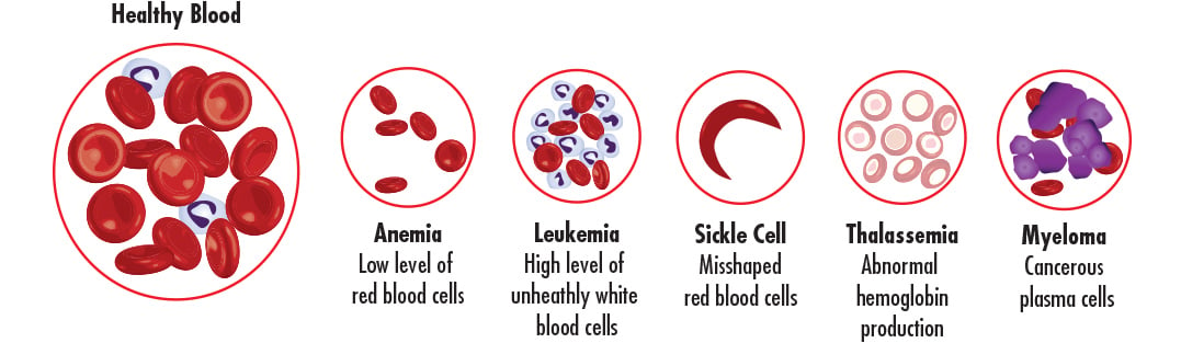
Known as blood cancer, leukemia is a malignant and progressive disease where bone marrow and other blood producing regions yield abnormal or premature leukocytes, which suppresses the production of normal, healthy blood cells in the blood plasmaThe primary liquid component of blood, often considered the vehicle or matrix that holds the red blood cells, white blood cells, and platelets together. Plasma is made up of roughly 95% water, which accounts for a little over half of humans’ total blood volume. The remaining bits of plasma are made up of a mixture of various proteins, sugars, clotting elements, and various electrolytes..
Result of various disorders that causes red blood cellsThe most common type of blood cell and primary oxygen transporter in the body. Red blood cells are transported by blood and the beating of the heart in the circulatory system. Red blood cells are primarily composed of a protein called hemoglobin and are produced by bone marrow inside of bones. to become misshapen or break down, along with other problems in the cardiovascular system.
Disorder of the blood with abnormal forms of hemoglobinIron-containing protein of red blood cells primarily responsible for oxygen transport throughout the body. This oxygen is delivered throughout the body to critical tissues and organs in order to complete the metabolic processes critical for the body to function. that can result in tiredness, bone problems, an enlarged spleen, yellowish skin, and slow growth among children.
Disorder of bone marrow that yields increased red blood cellsThe most common type of blood cell and primary oxygen transporter in the body. Red blood cells are transported by blood and the beating of the heart in the circulatory system. Red blood cells are primarily composed of a protein called hemoglobin and are produced by bone marrow inside of bones. counts and can also cause an increase in white blood cellsWhite blood cells, commonly referred to as leukocytes, are critical to our immune system and combat infectious diseases and bacteria. Most often found in blood and lymph nodes, white blood cells are derived from bone marrow. They contain a nucleus and are typically larger and denser than other cells and proteins in blood. The total count of white blood cells in the human body is often directly correlated to bodily health. and plateletsComponent of blood with a primary role of clotting and stopping blood flow at the site of an injury. Platelets are much smaller than red blood cells. Unlike white blood cells, platelets lack a nucleus, making them easily identifiable in a blood sample. .
Disorder in which blood does not clot normally, causing sufferers to bleed severely from even slight injuries.
Deficiency of red blood cellsThe most common type of blood cell and primary oxygen transporter in the body. Red blood cells are transported by blood and the beating of the heart in the circulatory system. Red blood cells are primarily composed of a protein called hemoglobin and are produced by bone marrow inside of bones. or hemoglobinIron-containing protein of red blood cells primarily responsible for oxygen transport throughout the body. This oxygen is delivered throughout the body to critical tissues and organs in order to complete the metabolic processes critical for the body to function. in the blood, resulting in exhaustion.
Cancer of plasma cells in blood which results in weakened bones.


or view regional numbers
QUOTE TOOL
enter stock numbers to begin
Copyright 2025 | Edmund Optics BV, De Maas 22B, 5684 PL Best, The Netherlands
California Consumer Privacy Acts (CCPA): Do Not Sell or Share My Personal Information
California Transparency in Supply Chains Act
This content may include material that has been generated or modified using artificial intelligence (AI).
The FUTURE Depends On Optics®