Engineers are available to assist.

Americas — $10,000 in Products
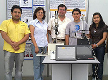
Universidad Peruana Cayetano Heredia, Peru — submitted by Mirko Zimic
For a low cost microscope capable of early detection of Tuberculosis built from stock optical components. The next generation microscope will enable cytopathology screening replacing the need of traditional Pap smear testing often unavailable to underprivileged women in remote or developing areas. Zimic's invention enables low cost, remote and fast telediagnostics of endemic diseases. His microscopes are already implemented in clinics in poverty stricken areas of Peru.
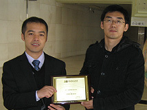
Asia — ¥800,000 in Products
Tohoku University (東北大学), Japan — submitted by Makoto Ohta
For contributing to the development of future medical instruments focused on understanding and monitoring the blood flow status in our bodies. Blood flow is a key to understanding the mechanism of why a cerebral aneurysm and stricture breaks out and grows. Ohta's invention is to represent the blood flow status faithfully in vitro and visualize the para-blood flow made of polymer gel by using PIV (Particle Image Velocimetry) technology. His research helps understand how to prevent a cerebral aneurysm and stricture in the future, helping to cure cardiovascular diseases, one of the leading causes of mortality!
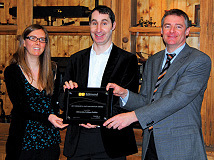
Europe — €5,000 in Products
Technische Universität Ilmenau, Germany — submitted by Stefan Sinzinger
Research focuses on the development of optical systems to improve techniques such as optical tweezing and digital holography for the micromanipulation and analysis of particle flows in integrated microfluidic systems. Highly integrated microfluidic systems are widely used for biomedical applications such as drug testing and disease treatments. Due to the high degree of miniaturization and integration, microfluidics enable investigations with extremely low probe volumes which is helpful for speeding up research and development in this field at reasonable cost. Both fluorescence detection and optical tweezers are extremely helpful in the handling and analysis of biological specimen. It is therefore of specific interest to adapt optical tweezers for the use in microfluidic systems.
Americas — $7,500 in Products
Rockefeller University, USA — submitted by Dan Gareau
For developing novel approaches to confocal microscopy for biological applications which focuses on noninvasive methods. One such example, in fiber optic spectroscopy, enables tissue oximetry (different than pulsed oximetry) that identifies ischemic injury as a flag for surgical intervention to prevent postoperative failure of surgically modified tissues. Additionally Gareau and his students, some as young as 14 years old, are involved in research programs incorporating students from grade school to graduate school including participants from: Oregon Health & Science University, Columbia University, The Bronx School of Science, Beaverton School of Science and Technology, and Moses Brown School.
Asia — RMB48,000 in Products
CIOMP (中科院长春光机所), China – 张 志军
Changchun Institute of Optics, Fine Mechanics and Physics, Chinese Academy of Sciences (CIOMP) is currently researching novel techniques for high power semiconductor lasers. Such applications being investigated include laser pumping, optical communication, laser display and medical treatments. The team has reached great achievements and requires additional optical components microscopy products and precision mechanics in order to advance this project.
Europe — €3,000 in Products
University College Dublin, Ireland — submitted by James Rice
This project involves development of an optical nanoscopy and nanoimaging tool, which will uniquely achieve sub-surface and sub-100 nm chemical mapping of biological (and functional) materials. The method combines AFM and optical techniques, with high spatial imaging resolution provided by the AFM scanning tip and will enable chemically specific information to be obtained via directly measuring infrared (IR) absorption spectra or 'fingerprint' to build an IR absorption image. This tool will address specific questions in regards to interactions that nanomaterials have within soft-condensed matter environments, at potentially the single nanoparticle or single molecule level, providing information not presently accessible using current imaging methodologies. Present IR absorption microscopy technology cannot image on the nanoscale. One of the areas of research where a limitation in the resolution in optical microscopy is significant is in nanobiology. An important area of research in nanobiology is the application of nanomaterials to biosystems which offers significant potential in a range of areas, for example in the development of new treatments, medicines and the creation of hybrid bio-electronic devices.

Americas — $5,000 in Products
University of Virginia, USA — submitted by Paul Yates
For his low cost, portable, easy to use, point and shoot high image quality retinal cameras for teleretinal screening for eye diseases such as diabetic retinopathy, glaucoma, and retinopathy in adults. This technology will be also used for vision screening of premature infants. Yates recently received a grant from State of Virginia to validate his technology nationwide and provide a case study to FDA. Early diagnosis of retinopathy is key in prevention of vision loss. The technology has already been commercialized and approved in Europe, and his cameras are currently being used in India and Thailand.
Asia — USD$5,000 in Products
University of New South Wales, Australia — submitted by Hans Riesen
University of New South Wales, Canberra is developing the most compact dosimetry system available globally. The system is based on patented X-ray storage phosphor technology and a laser based readout system involving sophisticated optical assembly and detector that can be incorporated into mobile phone sized housing. The system can monitor large numbers of people in radiation space during nuclear accidents such as the recent Fukushima event or after deployment of dirty bombs. It is also suitable for the decentralized and daily monitoring of medical personnel and other workers that may be exposed to ionizing radiation such as X-rays. This system will greatly benefit the general population in terms of radiation protection and allowing a significant reduction of radiation dose in medical imaging.
Europe — €2,000 in Products
Polytechnic University of Valencia, Spain — submitted by Verónica Sáiz
For research involving combining machine vision sensors and GPS for vegetative vigor mapping of small and midsized wine farms, determining, among others, the intensity of photosynthetic activity and consequently the plant health status. The project makes use of ground vehicle-mounted camera equipment in visible, UV, NIR mode for subsequent image analysis and mapping, while avoiding expensive and large-scale airborne data systems. This new approach introduces a non-invasive, less costly method of agricultural monitoring that can be applied to a variety of industries.
Americas Finalists
Asia Finalists
Europe Finalists


or view regional numbers
QUOTE TOOL
enter stock numbers to begin
Copyright 2025 | Edmund Optics BV, De Maas 22B, 5684 PL Best, The Netherlands
California Consumer Privacy Acts (CCPA): Do Not Sell or Share My Personal Information
California Transparency in Supply Chains Act
This content may include material that has been generated or modified using artificial intelligence (AI).
The FUTURE Depends On Optics®