Engineers are available to assist.

by Randall Hinton
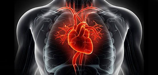
The future depends on fast and accurate feedback from arterial blood tests during ventilator monitoring to aid in recovery.
Medical staff carefully measure the amount, type, speed, and force of the air the ventilator pushes into and pulls out of a patient’s lungs and any complications that may arise. For example, too much oxygen in the blood for too long can be detrimental for the lungs. Also, too much acid in a patient’s blood can indicate kidney failure, a severe infection, specific toxic ingestions, diabetic ketoacidosis (DKA), or under-treated sleep apnea. Preventing these issues requires the frequent measurement of arterial blood gas levels.
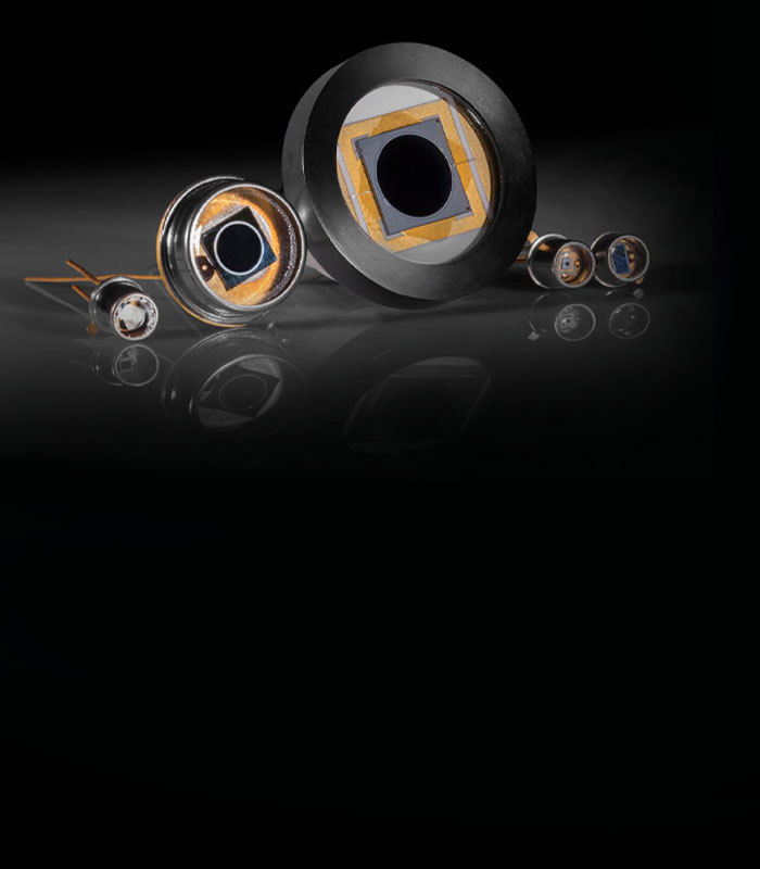
A blood sample is generally taken from the patient’s wrist, an artery in the groin, or the inside of the arm above the elbow8. The sample is then placed in a blood gas analyzer device. Blood gas analyzers measure the amount of oxygen (O2), carbon dioxide (CO2), and acidity of blood pH. They also measure concentrations of other parameters such as lactate, glucose, hemoglobin, creatinine, and electrolytes2. Examples of prominent companies that manufacture blood gas analyzers include Siemens Healthineers, Instrumentation Laboratory, Radiometer, Roche Diagnostics, and Nova Biomedical7.
Carbon dioxide, oxygen, pH, and hemoglobin levels are key parameters in arterial blood gas monitoring. Below are several examples of how those parameters may be photometrically determined.
Figure 1 shows an optical system for photometric determination of CO2 from a blood sample. Radiation from a thermic source is reflected from a concave mirror through a slit and onto a second concave mirror. The radiation is reflected from the second concave mirror to a grating, which is optimized for IR wavelengths from 4200-4300nm. The grating rotates, allowing the diffracted wavelengths to vary across this spectrum. The radiation is then transmitted back to the concave mirror, passed through a 3500nm longpass filter, transmitted through the measuring chamber, and then directed to a pyroelectrical detector. The detector, which is synchronized with the rotating grating, emits a signal proportional to the radiation intensity at 4210, 4260, and 4310nm. The amount of absorption from these wavelengths provides the baseline amount of CO2 in the sample3. Arterial blood should be relatively depleted of carbon dioxide because of pulmonary gas exchange, or external respiration4.

Figure 2 shows an optical system used for the determination of the amount of oxygen in a blood sample. Light from a green diode is transmitted through a 580nm short wave pass (SWP) filter. The radiation is transmitted from the SWP filter to a 580nm dichroic filter that reflects the green diode light onto the focus lens. The green light is then focused on the sample where it interacts with an oxygen-sensitive dye5. The excited luminophores emit photons with longer wavelengths than the incident green light. These excitation photons are then transmitted from the sample container through the lens, through the dichroic filter, then through a 665nm longpass color filter, and onto a silicon photodiode. The signal intensity at the detector is used to calculate the oxygen level. Oxygen levels are dependent on movement of air in and out of alveoli, blood flow through pulmonary capillaries, and oxygen carrying protein hemoglobin4.

Figure 3 shows a cross section of an optical system for photometric determination of pH. A halogen lamp emits radiation that is filtered by a heat absorbing filter, such as SCHOTT KG5 glass. The radiation then interacts with the blood sample and is finally focused by a lens through several bandpass filters onto silicon photodiodes. The photodiodes emit current signals representing the intensity of 458, 589, and 750nm radiation. These radiation intensities allow for the pH value of the sample to be calculated3. Blood pH provides insight into the interaction of chemical buffers present in blood (principally bicarbonate), red blood cells, and the function of three organs: the kidneys, lungs, and brain stem4.

Oxygen is poorly soluble in blood, so the body relies on the oxygen carrying protein, hemoglobin, to transport oxygen to tissue cells. An optical system known as a CO-oximeter measures the absorption of light passing through blood from as few as two or three wavelengths of light to several dozens of wavelengths. This is done in order to distinguish oxyhemoglobin and deoxyhemoglobin (formerly called "reduced" hemoglobin) and determine the oxyhemoglobin saturation, or the percentage of oxygenated hemoglobin compared to the total amount of available hemoglobin (Hb). Measuring greater numbers of wavelengths enables the instrument to distinguish between these and carboxyhemoglobin (COHb), methemoglobin (metHb), other hemoglobin moieties, and "background" light-absorbing species.

Figure 4 shows an example of a CO-oximeter. Radiation is transmitted from a halogen lamp through a heat absorbing filter onto the measuring chamber. The light that passes through the sample is focused through a dichroic filter, which then splits the spectrum into either a 600 or 506nm bandpass filter. The resulting intensities are detected by silicon photodies and used for the calculation of the total hemoglobin content (Hbtot) and oxygen saturation. The calculation is based on Lambert Beer's Law and predetermined values of the extinction coefficients for Hb and HbO2 3.
Below is another example of a CO-oximeter, where the red blood cells are exposed to ultrasonic vibration which destroys the cell walls releasing the hemoglobin. Hemoglobin in the flow cell is exposed to visible light and the absorbance spectrum (478-672nm) is observed using a grating mirror and photodiode array. From this absorbance spectra, bilirubin concentrations can be computed and used to screen for conditions such as neonatal jaundice6.


or view regional numbers
QUOTE TOOL
enter stock numbers to begin
Copyright 2025 | Edmund Optics BV, De Maas 22B, 5684 PL Best, The Netherlands
California Consumer Privacy Acts (CCPA): Do Not Sell or Share My Personal Information
California Transparency in Supply Chains Act
This content may include material that has been generated or modified using artificial intelligence (AI).
The FUTURE Depends On Optics®